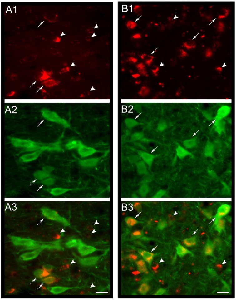
Figure 4.
MCH-rhodamine is internalized in DR serotonergic neurons. In (A1,B1), the photomicrographs shows that MCH-rhodamine (red) was internalized by DR neurons in the cat. Photomicrographs in (A2,B2) depict neurons with serotonin immunoreactivity (green). Superimposition of both photomicrographs is shown in (A3,B3). It is readily observed that MCH-rhodamine is present in serotonergic (arrows) and non-serotonergic neurons (arrowheads). Calibration bars: 20 μm. Orginal microphotographs taken from the data set of Devera et al. (2015).

