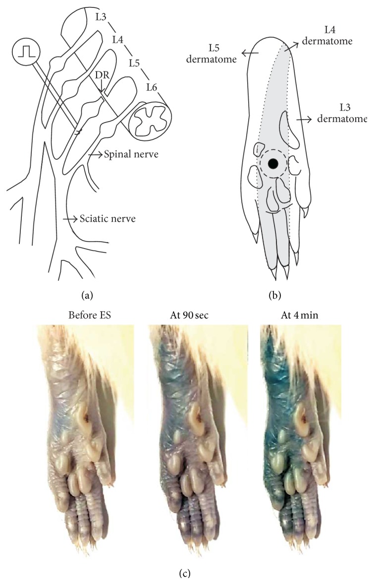
Figure 1.

The locations of electrical stimulation (ES), intraplantar (i.pl.) injection of substances, and paw sensitivity testing and an example of ES-induced plasma extravasation. (a) An L5 spinal nerve (SN) was decentralized by L5 dorsal rhizotomy (DR). A high-level tetanic ES (2–4 mA, 0.5 ms pulse, 4 Hz, and 5 min) was applied to the decentralized L5 SN 4–6 mm distal to the L5 dorsal root ganglion (DRG). (b) The tactile sensitivity testing was performed in an area (marked by a filled circle) located in the center of the circle surrounded by the tori, which is almost matched to the midpoint of the L4 plantar dermatome (shaded area) [29] of the hind-paw. A 30 μL i.pl. injection of different substances was administered subcutaneously into the skin area subjected to tactile sensitivity testing, forming a bleb that disappeared within 10 min after the injection (bleb area marked by a dotted circle). (c) Following Evans blue injection (i.v., 30 mg/kg, and 2% solution), areas of plasma extravasation induced by the ES of the L5 SN are seen as blue. Note that dye extravasation covered the L5 plantar dermatome within the first 90 seconds of the ES, and the extravasation area extended to the L4 plantar dermatome within 4 minutes of the ES.
