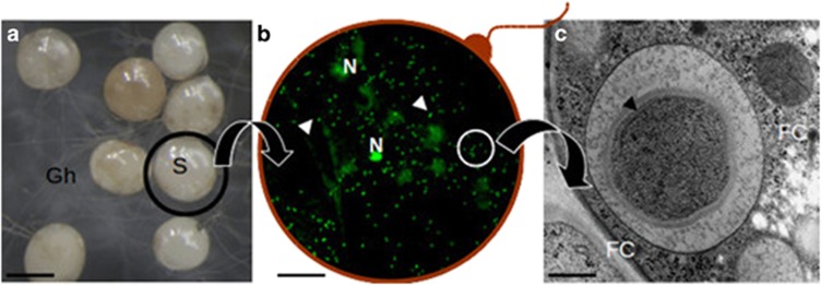
Figure 1.
A composite picture illustrating the location of CaGg. (a) G. margarita spores (S) observed under a stereomicroscope produce a network of germinating hyphae (Gh); (b) a squashed spore reveals the fungal nuclei (N) and a multitude of endobacteria (arrows) after staining with Bacteria Counting Kit component A and observation by confocal microscopy. The red line is drawn to suggest the fungal wall; (c) a bacterium observed by electron microscopy reveals the multilayered Gram-negative wall; it is located inside the fungal cytoplasm (FC), limited by a membrane of fungal origin. Bars correspond to 230 μm in a, 13 μm in b and 0.35 μm in c.

