
Fig. 2.
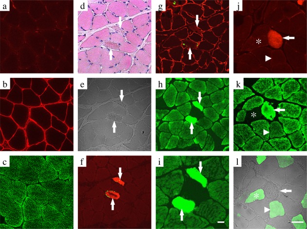
Conformity of cathepsin-D expression and apoptotic myofibers by immunohistochemical analysis in the soleus muscle of the CON (a–c) and HULT (d–l) groups. Transverse serial sections of the muscle were labeled with both anti-active caspase-3 antibody and TUNEL (a, f), both anti-dystrophin antibody and TUNEL (b, g), anti-cathepsin-D antibody (c, h, i, k), anti-active caspase-3 antibody (j), and fast myosin antibody (l). A higher magnification of panel e is depicted in panel i. Some of the serial sections were routinely stained with eosin–hematoxylin solution (d). Arrows indicate apoptotic myofibers expressing a positive reaction for both the active caspase-3 (red) and TUNEL-labeling (green) in serial sections (d–i) and a positive reaction for the active caspase-3 (red) in serial sections (j–l). Asterisks indicate type II myofibers exhibiting a negative reaction for both cathepsin-D (green) and positive reaction for fast myosin (green) in the serial sections (j–l). Arrowheads indicate a hybrid-type myofiber exhibiting a positive reaction for both cathepsin-D and fast myosin in the serial sections (j–l). Scale bar: 20 μm.