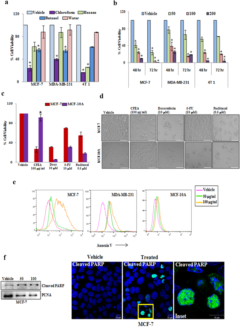
Figure 1. CFEA induces breast cancer cell selective cytotoxic effects and promotes apoptosis.
(a) MCF-7, MDA-MB-231 and 4T1 cells were treated with 100 μg/ml of different fractions (chloroform, hexane, n-butanol and water) of E. alba for 48 hours and cytotoxicity was measured by SRB assay as described in materials and methods. Percent cell viability were tabulated. Columns, average of triplicate readings of samples; error bars, ± S.D. *p < 0.01, compared to vehicle treated cells. (b) MCF-7, MDA-MB-231 and 4T1 cells were treated with increasing concentrations 50, 100, and 200 μg/ml of CFEA for 48 and 72 hours and cytotoxicity was assessed by SRB assay. Columns, average of triplicate readings of samples; error bars, ± S.D. *p < 0.01, compared to vehicle treated cells. (c) MCF-7 (red bar) and MCF 10A (violet bar) cells were treated with CFEA and FDA approved standard anti-cancer drugs Doxorubicin (10 μM or 5.79 μg/ml), Paclitaxel (0.5 μM or 0.42 μg/ml), and 5-Fluorouracil (10 μM or 1.3 μg/ml) for 48 hours and cytotoxicity was assessed by SRB assay. Columns, average of quadruplet readings of samples; error bars, ± S.D. *p < 0.01, compared to MCF 10A cells treated with different standard drugs. (d) Respective photomicrographs of vehicle and treated (48 hours) cells were shown. Scale bar, 100 μm. (e) Analysis of apoptosis induced by CFEA in MCF-7 (left panel), MDA-MB-231 (middle panel) and MCF 10A (right panel) cell lines. Cells treated with CFEA for 24 hours were stained with Annexin-V Alexafluor 488 and analysed by flowcytometry. (f ) Western blot analysis (left panel) of Cleaved PARP in nuclear fraction of 24 hours post vehicle and CFEA treated (50 and 100 μg/ml) MCF-7 cells. Vehicle or CFEA (100 μg/ml) treated (24 hours) cells were also stained with cleaved PARP antibody and analysed by confocal microscopy (right panel). Scale bar, 10 μm. Results shown from (a) to (f) sections are representative of at least three independent experiments.

