Figure 4.
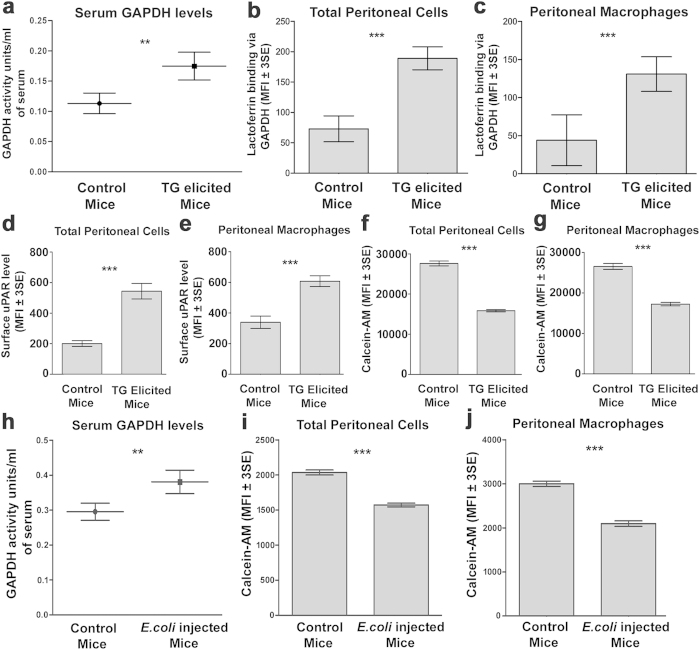
Peritoneal inflammation/infection enhances GAPDH secretion, lactoferrin capture and cellular iron absorption (a) TG elicited mice demonstrate enhanced serum GAPDH levels. Female Balb/c mice (8–10 weeks old) were injected i.p with 2.5 ml TG or with PBS (control mice). After 5 days serum was collected and serum GAPDH activity was assayed, p ≤ 0.01, n = 4 each group. (b,c) Cells from TG peritonitis induced mice demonstrate enhanced sGAPDH mediated Lf binding as compared to cells from control mice, p ≤ 0.0001, n = 104. (d,e) Cells from TG injected mice also demonstrate enhanced surface expression of uPAR, p ≤ 0.0001 n = 104. (f,g) Calcein quenching assay reveals that peritoneal cells from TG injected mice have higher levels of intracellular iron as compared to control cells, p ≤ 0.0001 n = 104. (h) E.coli infected mice also demonstrate elevated serum GAPDH levels. Eight to ten week old female Balb/c mice were injected i.p with mid log phase E.coli MG1655 (1 × 10⁴) in 500 μl PBS or with PBS alone (Control mice). After 2 days serum was collected and serum GAPDH activity measured, p ≤ 0.01, n = 4 for each group. (I,j) Host cells from E.coli injected mice show enhanced intracellular iron as compared to control cells, p ≤ 0.0001 n = 104. In all flow cytometry analysis the macrophage population was identified using anti F4/80 staining. All experiments were repeated three times and representative graphs are presented.

