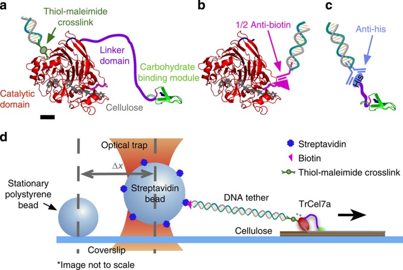
Figure 1. Constructs and assay schematic.
Construct details and optical trap assay schematic for (a) wtTrCel7A, where a DNA-bound sulfo-SMCC crosslinks through available surface lysines (scale bar, 1 nm), (b) isolated biotin-labelled CD ligated to DNA through a ½ anti-biotin antibody and (c) isolated CBM tethered through a DNA-bound anti-His antibody. Structures in a–c are from PDB 7CEL and 2CBH. (d) A schematic of the wtTrCel7A motility assay tracks motility through a 1,010-bp tether attached to a 1.25-μm streptavidin bead held in an optical trap. Stationary fiducial beads serve to compensate for drift.

