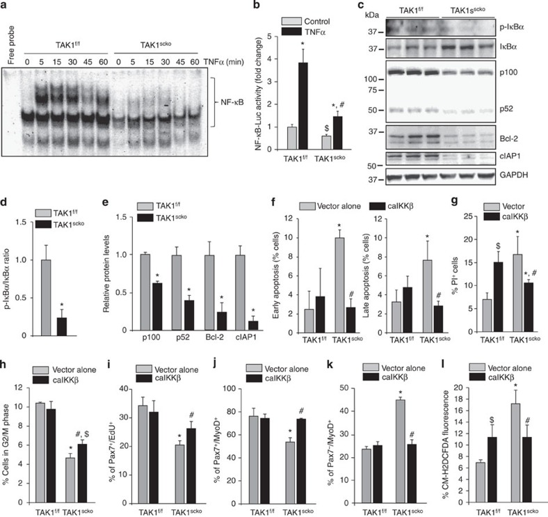
Figure 8. TAK1 is required for the activation of NF-κB in satellite cells.
(a) TAK1f/f and TAK1scko cells were treated with 10 ng ml−1 TNFα for indicated time period and the nuclear extracts made were analysed by performing electrophoretic mobility shift assay (EMSA). A representative EMSA gel is presented here (b) TAK1f/f and TAK1scko cells isolated from non-tamoxifen-treated mice were transfected with pNF-κB-Luc plasmid along with pRL-TK plasmid in 1:10 ratio for 24 h followed by treatment with 4-hydoxytamoxifen (TAM) for 48 h. The cells were then treated with 10 ng ml−1 TNFα for 18 h followed by measuring the amounts of luciferase and renilla activity in cell extracts using Dual luciferase assay kit. The fold change in luciferase/renilla ratio is presented here. N=3 in each group. *P<0.01, values significantly different from corresponding control TAK1f/f or TAK1scko cultures. #P<0.05, values significantly different from TNFα-treated TAK1scko cultures. $P<0.05, values significantly different from control TAK1f/f cultures. (c) Representative immunoblots presented here demonstrate the levels of p-IκBα, total IκBα, p100, p52, Bcl-2, cIAP1 and an unrelated protein GAPDH in TAK1f/f and TAK1scko cultures. (d) Densitometry analysis of ratio of phosphorylated and total IκBα protein in TAK1f/f and TAK1scko cultures. (e) Relative amounts of p100, p52, Bcl-2 and cIAP1 in TAK1f/f and TAK1scko cultures. TAK1f/f and TAK1scko cells isolated from non-tamoxifen-treated mice were transfected with vector alone or caIKKβ followed by treatment with 4-hydoxytamoxifen (TAM) for 48 h. The cells were then washed and incubated in growth medium and analysed at specific time points. Percentage of (f) apoptotic (AnnexinV+/PI+) (g) necroptotic (that is, PI+) cells in vector alone and caIKKβ-transfected TAK1f/f and TAK1scko cultures measured after 72 h of removal of TAM. Percentage of (h) cells in G2/M phase of cell cycle, (i) Pax7+/EdU+ cells, (j) Pax7+/MyoD+, (k) Pax7−/MyoD+ and (l) CM-H2DCFDA+ cells in vector alone or caIKKβ-transfected TAK1f/f and TAK1scko cultures measured after 48 or 72 h of removal of TAM. N=4 in each group for all the experiments in this figure. Error bars represent s.d. *P<0.01, values significantly different from corresponding TAK1f/f or TAK1scko cultures transfected with vector alone by paired t-test. #P<0.05, values significantly different from TAK1scko cultures transfected with vector alone by paired t-test. $P<0.05, values significantly different from TAK1f/f cultures transfected with vector alone by paired t-test.

