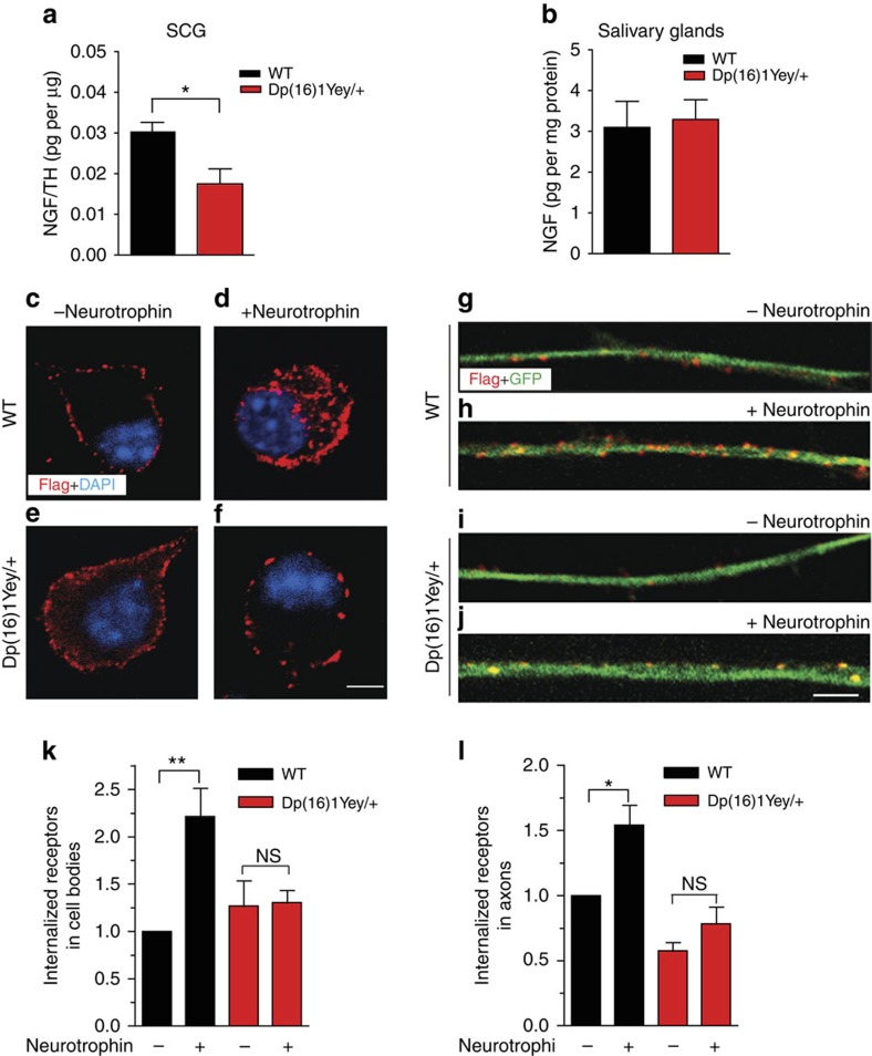
Figure 2. Impaired ligand-dependent endocytosis of Trk receptors in Dp(16)1Yey/+ neurons.
(a,b) Dp(16)1Yey/+ mice show a significant decrease in NGF protein levels in sympathetic cell bodies located in superior cervical ganglia (SCG; a), but not in the salivary glands (b), a target tissue innervated by sympathetic axons. NGF levels in SCGs were normalized to TH in SCGs and represented as picograms of NGF per μg of TH. NGF levels in salivary glands were normalized to total protein. Results are the mean±s.e.m. from n=3 Dp(16)1Yey/+ mice and four control litter-mates for SCGs, and n=5 mice per genotype for salivary glands. *P<0.05, unpaired two-tailed Student's t-test. (c–j) Ligand-dependent Trk receptor internalization is impaired in Dp(16)1Yey/+ sympathetic neurons. Sympathetic neurons from P0.5 Dp(16)1Yey/+ and wild-type (WT) sympathetic neurons were established in microfluidic chambers and infected with an adenovirus expressing FLAG-TrkB:A chimeric receptors. Neurons were labelled with FLAG antibody under non-permeabilizing conditions at 4 °C for 30 min, followed by BDNF treatment for 30 min. FLAG immunoreactivity (red) was assessed in cell bodies (c–f) and axons (g–j). Scale bars, 5 and 10 μm for axons and cell bodies, respectively. (k,l) Quantification of internalized Trk in cell bodies and axons after treatments described in c–j. Internal accumulation of chimeric receptors under the various conditions was determined by assessing the proportion of co-localization of FLAG immunofluorescence with that of GFP, which is co-expressed in infected neurons and is cytoplasmic. At least 40–50 neurons were analysed per condition. Quantification is represented as fold-change relative to WT neurons with no neurotrophin. Results are the mean±s.e.m. from three independent experiments. *P<0.05, **P<0.01, NS, not significant, two-way analysis of variance (ANOVA) followed by Bonferroni post-hoc test.

