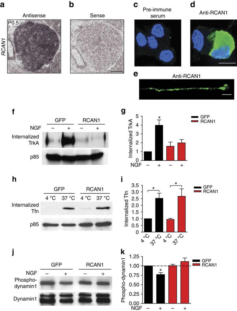
Figure 3. Increased expression of RCAN1, an endogenous calcineurin inhibitor, downregulates TrkA endocytosis by altering dynamin1 phosphorylation.
(a) In situ hybridization shows endogenous expression of RCAN1 mRNA in the developing mouse superior cervical ganglia at P0.5. Sense control is shown in b. Scale bar: 100 μm. (c–e) RCAN1 protein is localized to both cell bodies (d) and axons (e) of cultured sympathetic rat neurons as detected using a RCAN1 antibody. Staining with pre-immune serum control is shown in c. Scale bar, 10 μm for c,d and 5 μm for e. (f) A cell surface biotinylation assay shows that adenoviral overexpression of RCAN1.4 attenuates NGF-dependent TrkA internalization in cultured rat sympathetic neurons. Membrane proteins were subjected to cell-surface biotinylation. Internalized TrkA receptors were detected by surface stripping of biotin, neutravidin precipitation and TrkA immunoblotting. Supernatants were probed for p85 for normalization of protein amounts. (g) Densitometric quantification of internalized TrkA. Results are means±s.e.m. from four independent experiments. *P<0.05 significantly different from all other conditions. (h) Uptake of biotin-labelled transferrin (biotin-Tfn) is unaffected by RCAN1 overexpression in rat sympathetic neuron cultures. After internalization at 37 °C and acid washes to remove surface-bound transferrin, internalized biotin-Tfn was detected in neuronal lysates by neutravidin precipitation and immunoblotting using a transferrin antibody. Supernatants were probed for p85 for normalization of protein amounts. (i) Densitometric quantification of internalized biotin-Tfn. Results are means±s.e.m. from five independent experiments. *P<0.05 significantly different from corresponding controls at 4 °C. (j) NGF stimulation results in dephosphorylation of dynamin1, that is abrogated by excess RCAN1. Neuronal lysates were immunoblotted using a phospho-Ser778 dynamin antibody. Immunoblots were stripped and reprobed for total dynamin1 for normalization. (k) Densitometric quantification of phospho-dynamin1 levels normalized to total dynamin1 levels. All values are expressed relative to the no neurotrophin treatment in GFP-expressing neurons. Results are means±s.e.m. from seven independent experiments. *P<0.05 significantly different from all other conditions. Statistical analyses by two-way ANOVA and Bonferroni post-hoc test for g,i,k. Full-length blot scans are shown in Supplementary Fig. 8.

