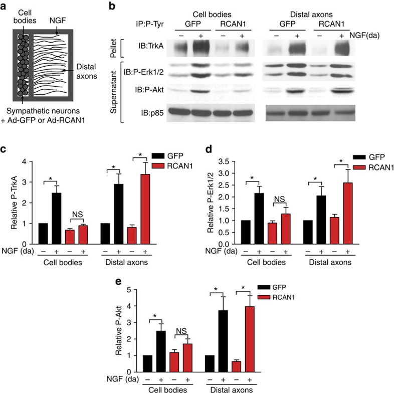
Figure 4. RCAN1 overexpression attenuates retrograde NGF signalling.
(a) Schematic of the compartmentalized culture system used for biochemical analyses of NGF-dependent signalling locally in distal axons and retrogradely in cell bodies. (b) NGF stimulation of distal axons promotes phosphorylation of P-TrkA, P-Erk1/2 and P-Akt locally in axons and retrogradely in cell bodies of control GFP-expressing sympathetic neurons. RCAN1 overexpression disrupts the propagation of a retrograde NGF signal to cell bodies but does not affect local activation of these effectors in distal axons. Distal axons (da) of sympathetic neurons expressing GFP or RCAN1 were stimulated with NGF (100 ng per ml) for 8 h. Cell body/proximal axon and distal axon lysates were prepared and subjected to immunoprecipitation with a P-Tyrosine (PY20) antibody followed by immunoblotting for TrkA to detect P-TrkA. Supernatants were immunoblotted for P-Erk1/2, P-Akt and p85. (c–e) Densitometric quantifications of levels of P-TrkA (c), P-Erk1/2 (d) and P-Akt (e). P-TrkA, P-Erk1/2 and P-Akt signals were all normalized to p85 levels. Results are means±s.e.m. from five independent experiments, and expressed relative to no neurotrophin conditions. NS, not significant, *P<0.05 by two-way ANOVA and Bonferroni test. Full-length blot scans are shown in Supplementary Fig. 8.

