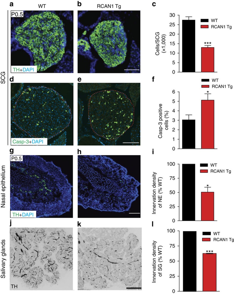
Figure 6. RCAN1 transgenic mice exhibit loss of sympathetic neurons and reduced sympathetic innervation of target tissues.
(a–c) Transgenic mice expressing three copies of RCAN1 (RCAN1 Tg) exhibit significant decreases in SCG size and cell numbers compared with litter-mate controls (WT) at P0.5. SCGs were visualized using TH immunohistochemistry and cell counts performed on Nissl-stained tissues. Values are the mean±s.e.m., n=5 mice each for wild-type and RCAN1 Tg mice. ***P<0.001. (d,e) Cleaved caspase-3 immunofluorescence shows enhanced apoptosis in P0.5 SCGs from RCAN1 Tg mice. (f) Quantification of percentage of SCG neurons that were immunoreactive for caspase-3. Values are the mean±s.e.m., n=5 mice for each genotype. *P<0.05. (g–l) TH immunostaining of sympathetic target tissues show substantial reductions in TH-positive sympathetic fibres within the nasal epithelium (g–i) and salivary glands (j–l) in RCAN1 Tg mice compared with litter-mate controls, at P0.5. For quantification of innervation density, the ratio of TH immunoreactivity to total image area was calculated from multiple images. The results are represented as a percentage of the mean for wild-type mice for nasal epithelium (i) and salivary glands (l). Values are the mean±s.e.m., n=3 mice for each genotype. *P<0.05, ***P<0.001. Statistical analyses by unpaired two-tailed Student's t-test. Scale bars, 100 μm.

