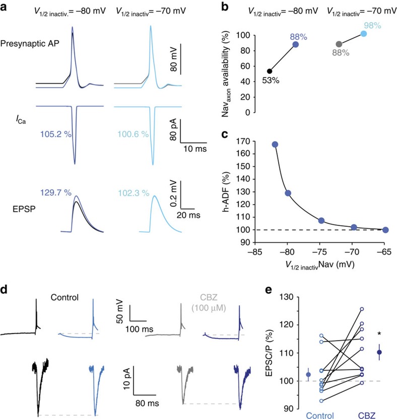
Figure 6. Role of Nav inactivation in h-ADF.
(a) Simulated h-ADF in control conditions (V1/2 inactivation=−80 mV for axonal sodium channels). Note the increased amplitude of the spike. Lack of h-ADF when the half-inactivation of the axonal sodium channel is depolarized (V1/2=−70 mV). (b) Summary of the availability of Navaxon with V1/2 inactivation=−80 mV or −70 mV. Note the marked increase with −80 but not −70 mV. (c) Magnitude of simulated h-ADF as a function of V1/2 inactivation of Nav channels in the axon. Note the increase in h-ADF induced by the hyperpolarization of V1/2. (d) Experimental enhancement of Nav inactivation with CBZ increases the magnitude of h-ADF. Under control condition (left), this connection expresses no h-ADF. When CBZ is added, h-ADF is now visible (right). (e) Quantitative data for 10 mature CA3–CA3 connections (DIV 24–32). Star: Wilcoxon, P<0.05.

