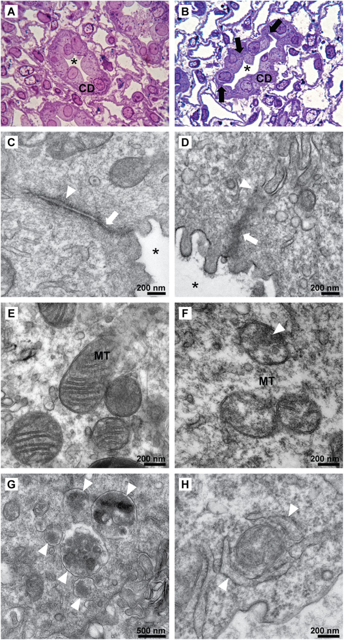
Figure 5. Structural abnormalities of IMCD in early potassium-deprived rats.
Compared to control IMCD (A), histopathology in potassium-deprived IMCD (B) shows disruption between IMCD cells (arrows) and a collapsed tubular lumen (*), Toluidine Blue, original magnification 400x. (C) An electron micrograph (EM) of the intercellular junction of control IMCD cells demonstrates a thin tight junction (arrow) and thin delicate adherens junction (arrow head). (D) A tight junction with disorganized filaments as well as thick electron dense deposition (arrow) and ill-defined adherens junction with thick electron dense deposition (arrow head) were observed in potassium-deprived IMCD cells. (E) Normal mitochondria in control IMCD cells. (F) Abnormal mitochondria in potassium-deprived IMCD show swollen cristae with the focal electron dense deposition (arrow head). Autophagolysosomes (G, arrow heads) and mitophagy (H, arrow heads) in IMCD cells of the potassium-deprived rats.

