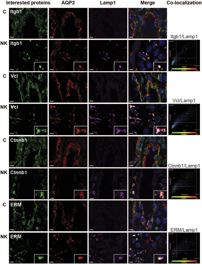
Figure 7. Co-localization of actin cytoskeletal and cell adhesion proteins with autophagy markers.
The inner medulla sections of control rats (C) and rats fed with a potassium-free diet for 1 day (NK) were triple-labeled against AQP2 (red), Lamp1 (pink), and down-regulated proteins including Itgb1, Vcl, Ctnnb1, and ERM (green). Insets demonstrate the areas where significant co-localization was observed as a group of puncta, which was not observed in control sections. Scale bar = 4 μm. Scatter plots demonstrate the degree of co-localization between lysosomal marker (Lamp1) and Itgb1 (r2 = 0.81, P < 0.05), Vcl (r2 = 0.54, P < 0.05), Ctnnb1 (r2 = 0.49, P < 0.05), or ERM (r2 = 0.42, P < 0.05) after potassium deprivation.

