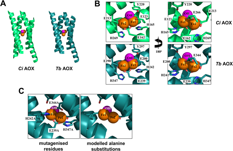
Figure 1. Structural modelling and mutagenesis of active site of Ciona intestinalis AOX.
(A) Model of the active site of the Ciona (Ci) enzyme, green, compared with the structure of the Trypanosoma brucei (Tb) AOX, blue. In both cases, the diiron site (iron moieties in orange, hydroxyl in pink) is buried in a four alpha-helix bundle. For clarity, only one protomer is shown. (B) Conserved residues binding the diiron centre show an identical arrangement in the Ci model (green) as in the Tb structure (blue). (C) The residues selected for alanine-substitution mutagenesis in the Ci enzyme (here shown in blue), alongside the resulting modelled structure.

