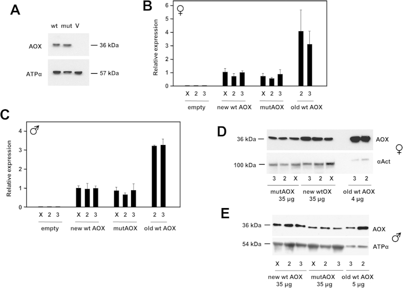
Figure 2. Expression of AOX transgenes in mammalian cells and Drosophila.
(A) Western blot of protein extracts from HEK293T cells transfected with wild-type and mutated AOX constructs (wt, mut) or empty vector (V), probed for AOX and for ATP synthase subunit α as loading control. (B,C) Relative AOX expression at RNA level, based on QRTPCR, in (B) females and (C) males of different Drosophila lines transgenic for wild-type or mutated AOX, or empty vector, inserted on chromosomes X, 2 and 3, as shown, in combination with the ubiquitous da-GAL4 driver. New wt (wild-type) and mutAOX lines were those created by site-specific integration at defined chromosomal sites using the ΦC31 system; old wt AOX lines were UAS-AOXF6 (chromosome 2) and UAS-AOXF24 (chromosome 3). For males, all values were significantly different from empty-vector lines; old wt AOX lines were significantly different from new wt AOX lines (p < 0.001, ANOVA followed by post-hoc Bonferoni-corrected t test), but mutAOX and new wt AOX lines were not significantly different from each other. Statistical analysis for females gave similar results, although greater sample-to-sample variation for old wt AOX lines yielded only p < 0.05 comparing them with new wt or mutAOX lines. (D,E) Western blot of protein extracts from the same flies (amounts as shown), probed for AOX or, as loading control, either ATP synthase subunit α or α-actinin, as indicated.

