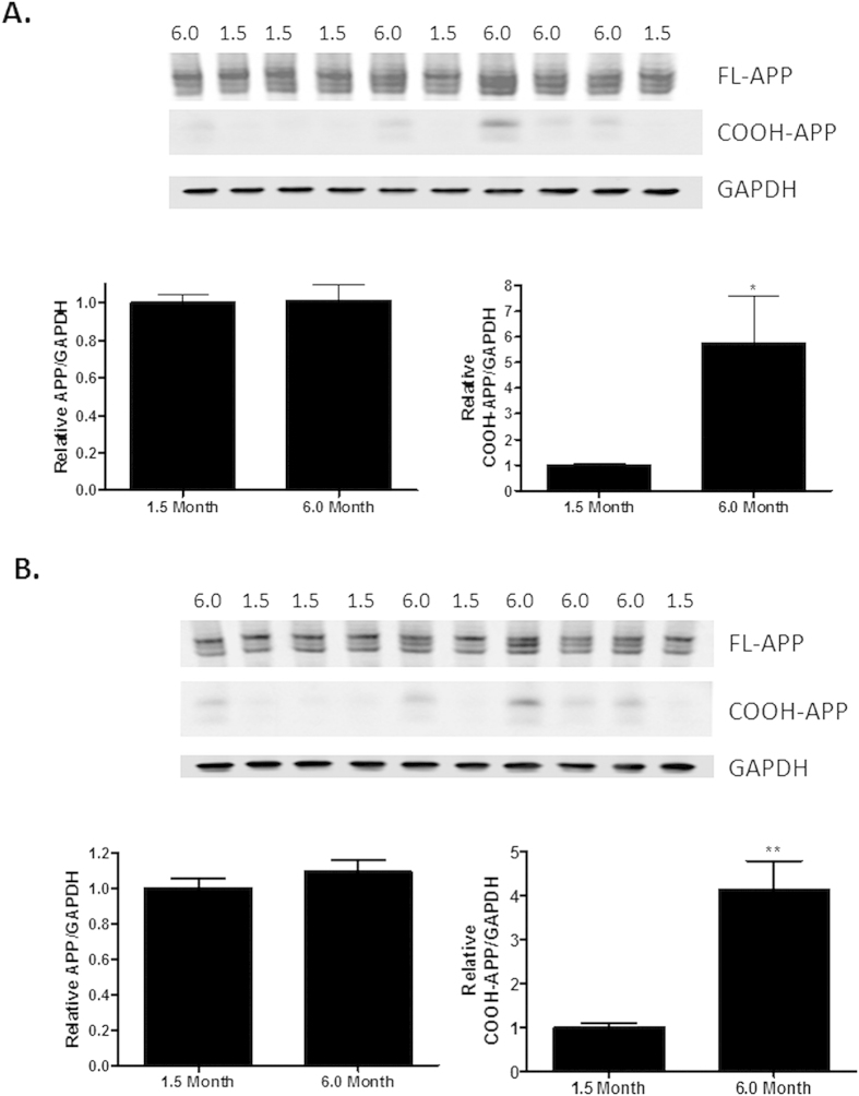
Figure 7. COOH-terminal APP antibody (5685) reveals increased COOH-terminal APP fragments, but not intact APP, in aged 5XFAD mice.
APP expression in (A) cortical and (B) hippocampal brain homogenates from 1.5 month old and 6.0 month old 5XFAD mice (same samples as in Fig. 6) was determined by immunoblotting with the COOH-terminal 5685 APP antibody after protein separation on a 4–12% gradient gel, with normalization to GAPDH. Lane loading in the immunoblot was as in Fig. 6. Statistical analyses consisted of a two-tailed t-test N = 5/group; *p < 0.05; **p < 0.01.

