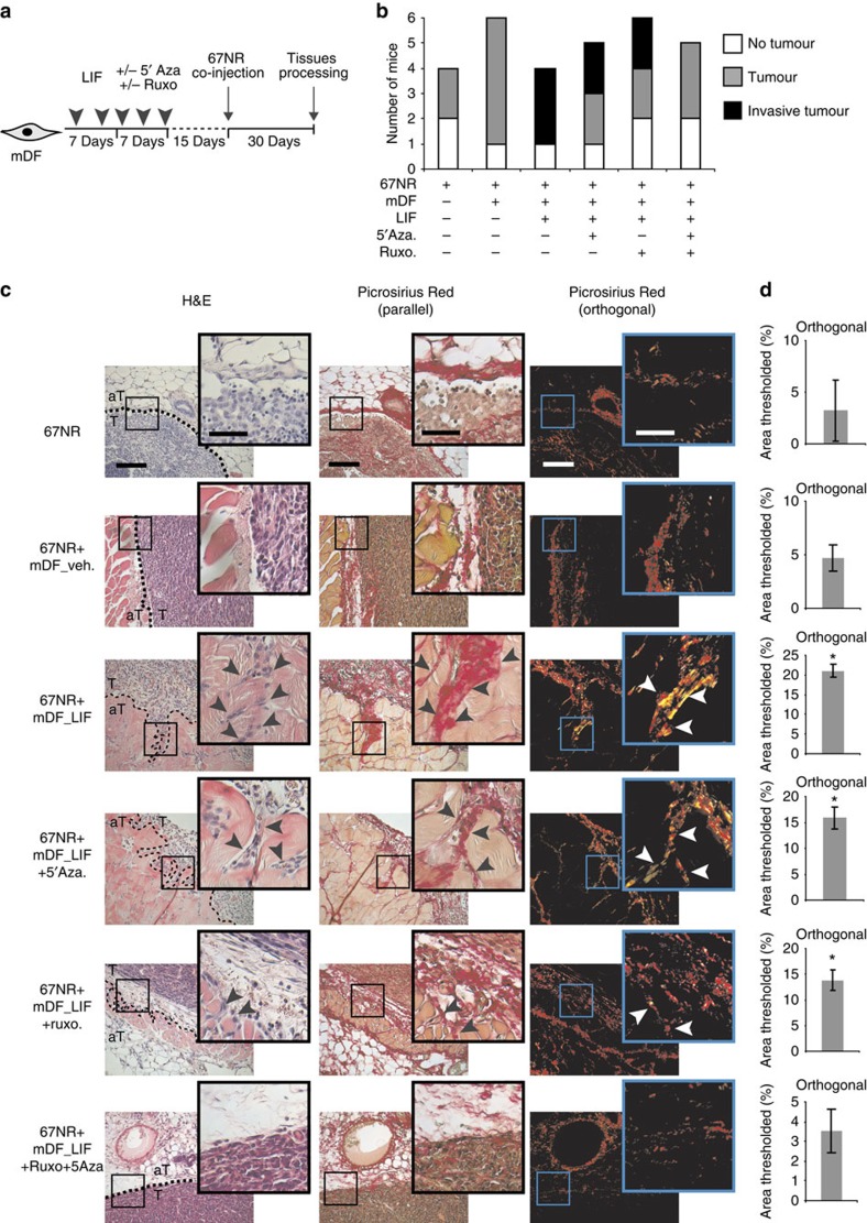
Figure 6. Stromal DNMT and JAK favour tumour invasion in vivo.
(a) Schematic representation of the experimental conditions used for in vivo cell co-implantation into the mammary fat pad of 8-week-old BALB/c female mice. 67NR mouse breast carcinoma cells were co-injected together with LIF preactivated mDF and treated with inhibitors. (b) Graphic representation of tumour formation induced by 67NR cells alone or in the presence of LIF-preactivated mDF. Total numbers of mice bearing tumours after single or combined inhibitor treatment are shown. (c) H&E coloration of paraffin-embedded sections of primary tumours isolated from mice (left panels) showing 67NR cells invading from the primary tumour (T) into the adjacent tissue (aT) (black arrow). Picrosirius Red staining visualized by both parallel (middle panels) and orthogonal (right panels) light showing tumour ECM remodelling at the areas invaded by the tumoral 67NR cells. Scale bars, 200 and 100 μm for × 20 and × 40X magnifications, respectively. (d) Quantification of Picrosirius Red staining using ImageJ software. Percentage of threshold area is shown (mean±s.d.; *P<0.05).

