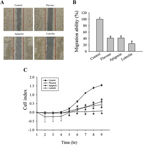
Fig. 3.

Flavone, apigenin and luteolin inhibited cell motility. a Representative images showing wound healing assays for cells treated with flavone (88 μM), apigenin (30 μM) or luteolin (43 μM) and an untreated control for 24 h. b Average number of cells that had migrated after 24 h. c Effects of the flavone, apigenin, and luteolin on MCF-7 cells migration. MCF-7 cells were treated with the IC50 concentrations (Table 1) of flavone, apigenin, and luteolin, and the real-time migration of the cells was measured using an xCELLigence system. The value of the open area at 0 h is 100 %. Results are the mean ± standard deviation of three independent experiments. P < 0.05 is considered as statistically significant. Symbols: *: P < 0.05; #: P < 0.01; ★: P < 0.001
