Fig. 1.

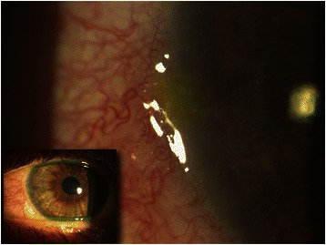
Digital image of the right eye (small picture). Limbal congestion with hyperemia and fluorescein staining of an epithelial defect overlying a marginal infiltrate at 9 o’clock limbus (in magnification)

Digital image of the right eye (small picture). Limbal congestion with hyperemia and fluorescein staining of an epithelial defect overlying a marginal infiltrate at 9 o’clock limbus (in magnification)