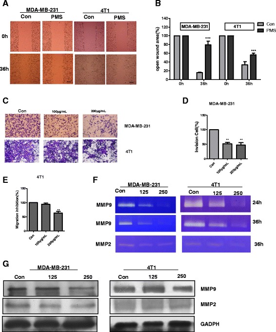
Fig. 3.

PMS inhibits migration and invasion of MDA-MB-231 or 4T1 cells by decreasing the activity of MMP insdead of the expression of MMP. a and b Effect of PMS on cellular migration by wound assay. a Confluent monolayers of cells were culture with solvent and with 200 μg/ mL PMS and the migration was evaluated by wound assay at 36 h. Scale bar = 100 μm. b The analysis of % open wound area was performed by the Tscratch software corresponding to the images in a. Data represent the mean ± S.D. of three independent experiments. c–e Effect of PMS on cellular invasion by transwell assay. c Cells were cultured with solvent or with 100, 200 μg/mL PMS onto the upper well coated with Matrigel. After 36 h treatment, cells passed though the Matrigel into the lower well were stained and counted. Scale bar = 25 μm (d) and (e) Analysis the % of invasion in comparison with control cell(100 %) corresponding to the images in c. Data represent the mean ± S.D. of three independent experiments. f PMS inhibits the activity of MMP9 and MMP2 secreted by MDA-MB-231 and 4T1 cells in vitro. The effect of PMS on MMP9 activity was tested by in gel zymography assay. Cells were cultured onto 6-well plates with solvent and with 125, 250 μg/mL PMS. After 24 h and/or 36 h treatment, cell supernatant was collected and performed Zymography. g Western Blotting showing the expression of MMP9 and MMP2 after 36 h treatment with solvent or 125, 250 μg/mL PMS. Similar results were obtained from independent experiments. The ** indicates significance different. The *** indicates extremely significance different
