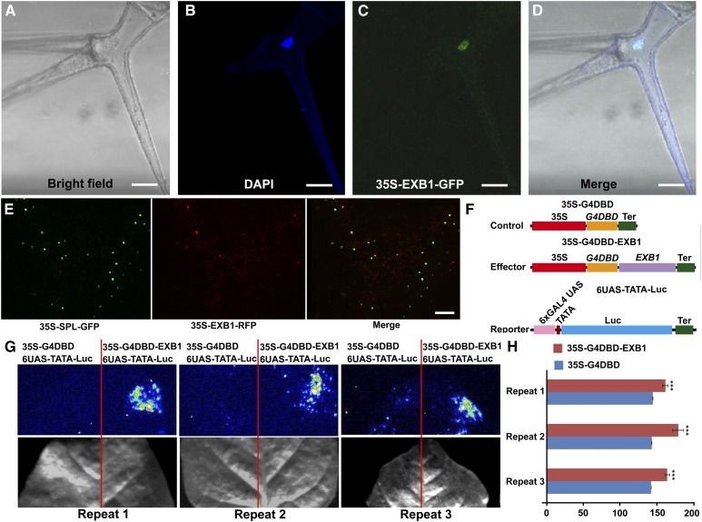
Figure 3.
EXB1 Is a Transcriptional Activator Localized in the Nuclei.
(A) to (D) EXB1 is localized in the nucleus. The leaf trichome cell of 35S-EXB1-GFP-5 transgenic line was observed. From left to right, the trichome under bright field, DAPI staining of the nucleus, GFP fluorescence, and merge of DAPI staining and GFP fluorescence. Bars = 25 µm.
(E) Coexpression of 35-EXB1-RFP and 35S-GFP-SPL in tobacco leaves indicated that EXB1 were colocalized with SPL, a known nuclear transcriptional repressor (Yang et al., 1999; Wei et al., 2015). Bar = 100 µm.
(F) to (H) EXB1 is a transcription activator.
(F) Schematic representation of constructs using in transcriptional activity assay.
(G) and (H) Transcription activity of EXB1 was tested in tobacco leaves using a GAL4/UAS-based system. The quantitative analysis of fluorescence intensity in (G) is shown in (H). Two-tailed t test was used to test the significance. Three asterisks represent P < 0.001. 35S, CaMV 35S promoter; G4DBD, GAL4 DNA binding domain; Ter, terminator of nopaline synthase gene; 6XGAL4 UAS, six copies of GAL4 binding site UAS; TATA, TATA box of CaMV 35S promoter.

