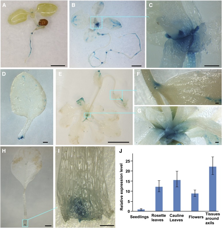
Figure 4.
EXB1 Is Expressed in Leaf Axils.
(A) The 7-d-old pEXB1-GUS-11 seedling. EXB1 was strongly expressed at the base of small true leaves.
(B) The 15-d-old pEXB1-GUS-11 plant. EXB1 was strongly expressed around the axils of rosette leaves.
(C) Close-up view of the leaf axils in the cyan square in (B).
(D) One leaf dissected from the plant in (B) showed that clear and specific expression of EXB1 at the leaf base.
(E) The 35-d-old pEXB1-GUS-11 plant. EXB1 was expressed in the axils of rosette leaves and cauline leaves.
(F) Close-up view of a cauline leaf axil in the cyan square in (E).
(G) Close-up view of the rosette leaf axils in the cyan square in (E).
(H) One leaf dissected from the plant in (E) showed a specific GUS staining at the leaf base.
(I) Close-up view of the leaf base in the cyan square in (H). EXB1 was specifically expressed at the position of AM initiation site.
(J) The relative expression of EXB1 in different tissues. EXB1 was expressed at the highest level in the tissues around axils. The expression level in seedlings was set to 1.0. The error bars represent the sd of three biological replicates.
Bars = 1 mm in (A), (B), and (H), 200 µm in (C), (D), (F), (G), and (I), and 1 cm in (E).

