Figure 6.
Differential Labeling of Microtubules by MAP65-1 and AIR9.
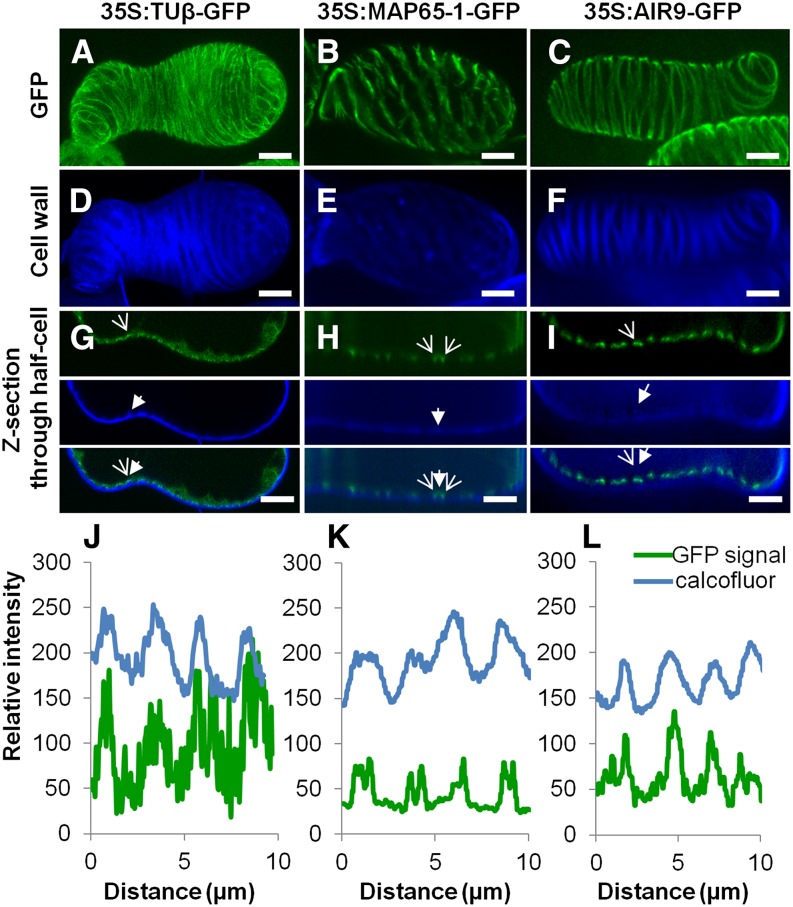
(A) to (C) Subcellular localization in TEs of tubulinß8 (A), MAP65-1 (B), and AIR9 (C) using constitutively expressed 35S promoter N-terminal GFP fusion proteins; bars = 5µm.
(D) to (F) Corresponding TE secondary cell wall stained with Calcofluor. bars = 5µm.
(G) to (I) Confocal cross section illustrating spatial relationship between GFP fusion protein and TE secondary thickenings for tubulinß8 (G), MAP65-1 (H), and AIR9 (I). Open arrows indicate fusion protein while closed arrows indicate TE cell wall thickening. Bars = 5 µm.
(J) to (L) Analysis of fluorescence intensity (8-bit values) distribution along the cell cortex (in µm) between Calcofluor-stained wall fluorescence and GFP fusion proteins. Note that tubulin accumulates mostly underneath TE thickenings (J). MAP65-1 flanks thickenings (K), whereas AIR9 accumulates exclusively beneath TE thickenings (L). Supplemental Movie 5 illustrates the subcellular localization of tubulin β8, AIR9, and MAP65-1 in TEs.

