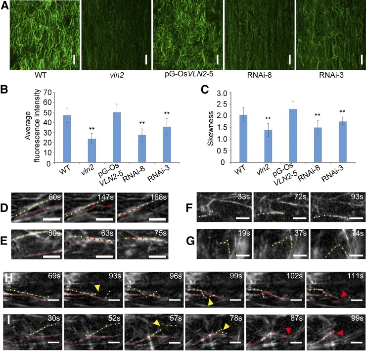
Figure 5.
The Organization and Dynamics of the Actin Cytoskeleton Are Altered in vln2.
(A) Staining of MFs in the root elongation zone by Alexa-488-Phalloidin in the wild type, vln2, a complemented line (pG-OsVLN2-5), and two RNA lines (RNAi-3 and -8). Images shown are the maximum projections of all optical sections (0.4-μm interval). Bars = 20 μm.
(B) Average fluorescence intensity of MFs in the wild type, vln2, pG-OsVLN2-5, RNAi-3, and RNAi-8, analyzed by MBF-ImageJ software. Five cells were analyzed and averaged in each root and five to eight roots were used as replicates for each of the lines.
(C) Skewness was measured to determine the bundling status of MFs in the wild type, vln2, pG-OsVLN2-5, RNAi-3, and RNAi-8. Five cells were analyzed and averaged in each root and five roots were used as replicates for each of the lines.
(D) to (I) Recording of MF severing, elongating, and bundling events by VAEM, based on fABD2-GFP decoration.
(D) and (E) Actin filament bundling events in the wild type (D) and vln2 (E).
(F) and (G) Actin filament elongating events in the wild type (F) and vln2 (G).
(H) and (I) Actin filament severing events in the wild type (H) and vln2 (I).
In (B) and (C), asterisks indicate significant difference from the wild type by Student’s t test, P < 0.01, and error bars indicate ±se. In (D) to (I), bars = 5 μm; yellow and red dotted lines indicate two adjacent MFs; colored triangles indicate the place where severing events occur.

