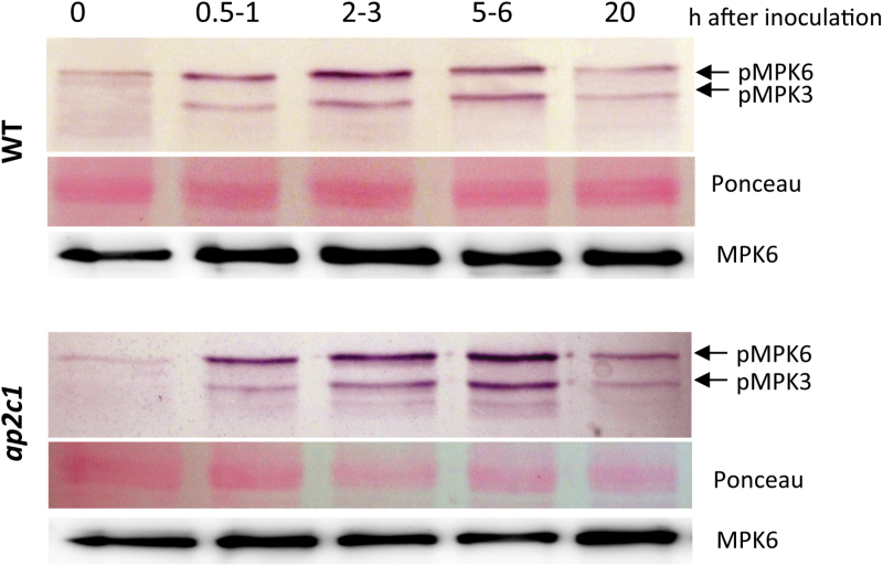
Fig. 1.
Analysis of MAPKs activation in roots during the early stage of H. schachtii infection. Phosphorylation of MPK6 and MPK3 was detected by immunoblotting with the anti-phospho ERK1/2 antibody. MPK6 protein amounts were detected with an MPK6-specific antibody. Ponceau-stained membranes present protein loading.

