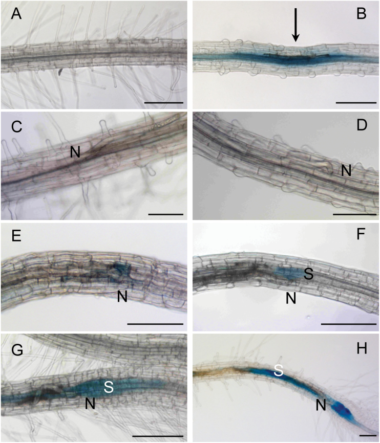
Fig. 4.
Microscopic analysis of AP2C1 promoter activity during H. schachtii infection in roots. AP2C1 promoter activity in (A) untreated pAP2C1::GUS root, (B) 1h after mechanical wounding (arrow points at the place of needle application), (C) during migration stage 2h after nematode infection (hai), (D) 4h after syncytium induction (hasi), (E) 15 hasi, (F) 24 hasi, (G) 30 hasi, and (H) 48 hasi. S, syncytium, N, head of the nematode. Bars: 200 μm.

