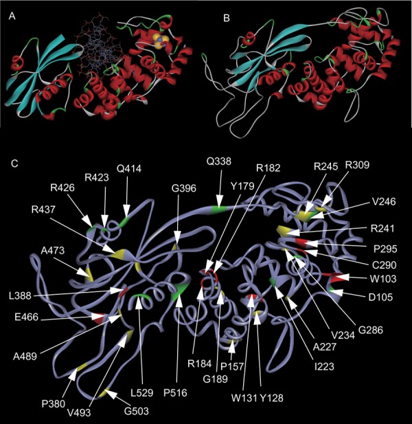
Figure 4.

Mapping of MUTYH variants on protein structure. A: Structure of B. stearothermophilus MutY bound to DNA. The iron and sulfur molecules in the [4Fe-4S] cluster are shown as yellow and gray spheres, respectively. B: Structure of MUTYH simulated by homology modeling. C: Mapping of amino acid residues examined in this study and functional information. Red sites show the residues with defective substitution. Yellow sites show the residues with partially defective substitution. Green sites show the residues with retained substitutions. p.R245, p.P405, p.R437, and p.A473 are also highlighted in yellow, as the residues with multiple substitutions that are defective and retained.
