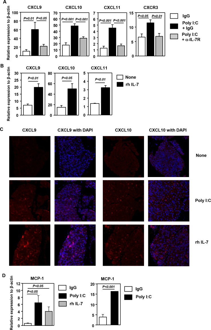
Figure 2.
Induction of CXCR3 ligands in the LAC by poly I:C and IL-7 treatment. (A) Real-time PCR analysis of mRNA levels of CXCR3 and its ligands in the LAC from C57BL/6 mice 24 hours after injection of poly I:C together with a blocking anti–IL-7Rα antibody or its isotype control IgG. The results are presented relative to that of β-actin (n = 6). (B) Real-time PCR analysis of mRNA levels of CXCR3 and its ligands in the LAC from C57BL/6 mice 24 hours after rh IL-7 injection, presented relative to that of β-actin (n = 3). Data are representative of four independent experiments. (C) Immunofluorescence staining of CXCL9 in the LAC sections from the mice treated with poly I:C described above. Data are representative of analyses of 10 mice each group (2–3 mice per experiment for a total of four independent experiments). (D) Expression level of MCP-1 in the LAC from C57BL/6 mice 24 hours after injection of poly I:C or rh IL-7 (left) or from mice 6 hours after injection of poly I:C (right), presented relative to that of β-actin (n = 4). Data are representative of three independent experiments.

