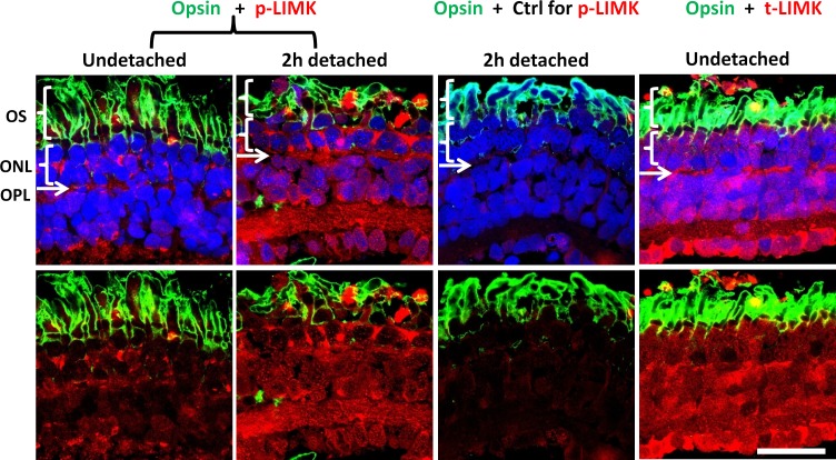
Figure 3.
Confocal microscopy demonstrates distribution of LIMK in salamander retina. The outer segments (OS) of rod photoreceptors are labeled for opsin (green). The outer nuclear layer (ONL) contains the photoreceptor cell bodies; nuclei are labeled with propidium iodide (blue). The outer plexiform layer (OPL) is located immediately beneath the ONL as a thin layer where nuclei are absent. Normal and 2-hour detached retinas, in the first and second columns, respectively, are labeled for p-LIMK (red); third column, a labeling control, anti–p-LIMK is omitted in 2-hour detached retina; fourth column, normal retina labeled for t-LIMK (red). Labeling of t-LIMK occurs throughout the normal undetached retina; p-LIMK signal is present but spotty in the normal retina and more diffuse, with an apparent increase, in 2-hour detached retina. Optical sections, 1 μm. Scale bar: 50 μm. n = 3 animals, 2 retinal explants per animal (retina from 1 eye was detached and cultured for 2 hours, retina from other eye was not detached and fixed immediately); 9 cryosections per group (3 cryosections per retinal explant), 2 or 3 images per cryosection.

