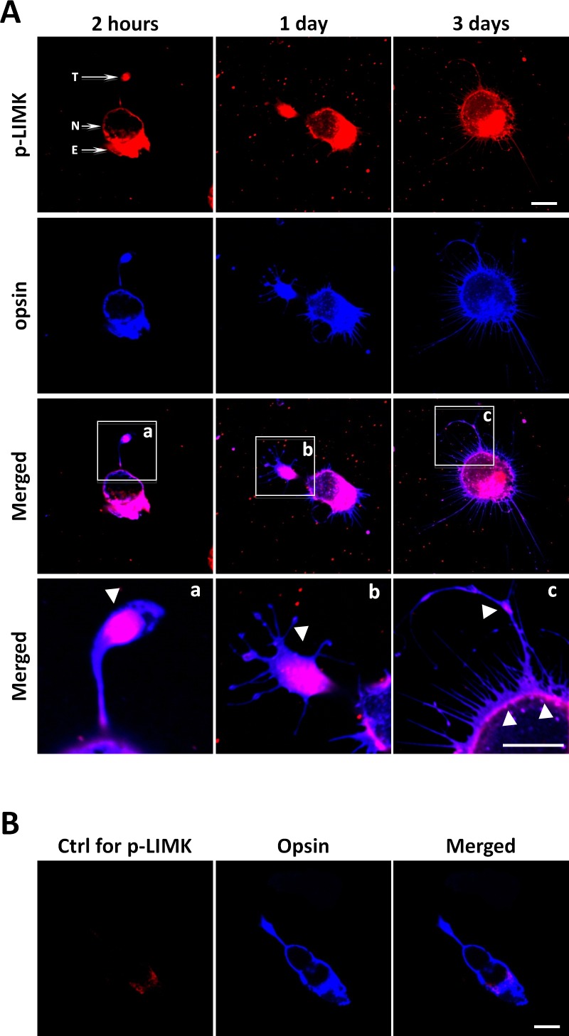
Figure 4.
Active LIMK is present in isolated salamander rod cells in culture. (A) Rod photoreceptors, which had lost their outer segments, double labeled for p-LIMK (red) and rod opsin (blue) and examined with confocal microscopy. Labeling occurs throughout the cell except in the nucleus, both during retraction, which occurs during day 1, and after growth of new processes, seen at 3 days. (a–c) Enlarged images show the presence of opsin in the cell membrane and p-LIMK in the cytosol and directly under the plasmalemma (arrowheads). (B) Control for p-LIMK labeling. Rod photoreceptor in 2-hour culture labeled with anti-opsin but without anti–p-LIMK, T, axon terminal; N, nucleus; E, ellipsoid, a collection of mitochondria, in the inner segment. Optical sections, 1 μm. Scale bars: 10 μm. n = 3 animals, 3 or 4 cultures per animal, 10 to 15 cells imaged per dish (∼39 cells per group).

