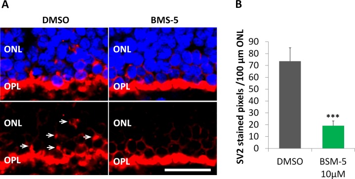
Figure 9.
Inhibition of LIMK reduces axonal retraction in the porcine retina maintained in vitro for 24 hours after detachment. (A) Representative control and treated retinas after 24 hours in vitro. The outer plexiform layer (OPL) with rod synaptic terminals is labeled for synaptic vesicles, SV2 (red). The outer nuclear layer (ONL) contains the photoreceptor cell bodies; nuclei are labeled with propidium iodide (blue). Left: With no treatment, SV2 label is present among the photoreceptor cell bodies (arrows) indicating that the rod terminals have retracted. Right: Treatment with 10 μM BMS-5, a LIMK inhibitor, reduced the level of SV2 in the ONL indicating an inhibition of retraction. Scale bar: 10 μm. (B) Retraction is measured using the area of SV2 label in the ONL. Treatment with 10 μM BMS-5 reduced SV2 labeling in the ONL significantly, compared to untreated retinas. Optical sections, 1 μm. n = 3 animals, 2 retinal explants per animal (1 explant with BMS-5 treatment from one eye, the other explant with DMSO as control from the other eye); 7 cryosections per group (2 or 3 cryosections per retinal explant), 3 or 4 images per cryosection. ***P < 0.001; Student's t-test.

