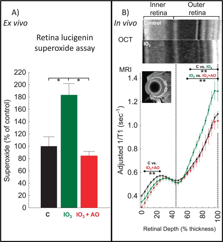
Figure 1.
Quench-assisted MRI measurement in vivo of outer retina oxidative stress. (A) Retina superoxide production measured ex vivo from dark-adapted controls (C, black, n = 10); sodium iodate–treated mice (IO3, green, n = 6); IO3 mice treated with AO (IO3+AO, red, n = 6). *Significant difference (P < 0.05). (B) Quench-assisted MRI profiles measured in vivo from dark-adapted controls (black, n = 30); IO3 mice (green, n = 7); IO3+AO mice (red, n = 9). Optical coherence tomography images (control vs. IO3) show mostly unchanged laminar spacing within the retina; dashed vertical lines map outer plexiform layer (48%) and retina/choroid boundary (100%) onto MRI profiles; MRI insert shows regions studied (white boxes); visual inspection of each group's MRI does not allow for easy appreciation of differences in the derived parameter 1/T1 and so only a representative image is presented. **Retinal depth range with significant difference (P < 0.05). Adjusted 1/T1 data at each depth used factors that normalize same-day controls to a control reference data set.

