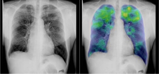
FIGURE 2.

CAD4TB image analysis. An image classified as 4 (highly suggestive of active PTB) by the expert consensus. Raw image on the left side and computer-generated overlay (i.e., the output of lung parenchyma texture analysis subsystem)19 of the same image on the right side. Note the highlighting (yellow/red) of the parenchymal lesions in the apical zones. CAD4TB abnormality score 87 (out of 100). CAD4TB = Computer Aided Detection for Tuberculosis; PTB = pulmonary tuberculosis.
