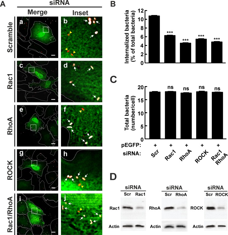
Fig 5. Knockdown of Rho GTPases and Rock inhibits internalization of C. burnetii.
(A) HeLa cells were co-transfected with pEGFP and a scramble (panels a and b), Rac1 (panels c and d), RhoA (panels e and f) or ROCK (panels g and h) siRNAs or the RhoA/Rac1 siRNA combination (panels i and j). Cells were infected for 4 h at 37°C with C. burnetii and then fixed and processed for immunofluorescence to determine C. burnetii internalization as described in Materials and Methods. Cells were analyzed by confocal microscopy. Representative micrographs of cells are presented. As indicated in Fig 1, in the merged images (panels a, c, e, g, and i) and the insets of the merged images (panels d, d, f, h, and j), extracellular C. burnetii is shown in white and red pseudo colors (arrows), while intracellular C. burnetii is shown in red pseudo color (yellow arrowheads). Scale bar: 5 μm. (B) Quantification of C. burnetii internalized by transfected HeLa cells. (C) Quantification of total C. burnetii associated to HeLa cells. Between 40 and 60 cells and between 400 and 600 bacteria were counted in each experiment. Results are expressed as means ± SE of three independent experiments. ***p < 0.001, compared to scramble siRNA (one-way ANOVA and Dunnett's post hoc test). ns: non-significant differences between groups (p > 0.05). (D) Lysates of cotransfected HeLa cells were analyzed by SDS-PAGE and Western blot using antibodies against Rac1, RhoA and ROCK. An anti-actin antibody was used as loading control. Scr: scramble siRNA.

