. 2015 Oct 5;34(22):2789–2803. doi: 10.15252/embj.201591320

Figure 4. Methylation of H3K9 spreads across natural boundaries and heterochromatin becomes derepressed in swi6∆ cells.

Figure 4

  1. H3K9me2 ChIP‐seq enrichment profiles for swi6+ and swi6∆ cells on centromere 1. Centromeric repeat elements (dg/dh) are indicated. Inverted repeats (IRC1) constitute heterochromatin boundaries in wild‐type cells (Cam et al, 2005). The y‐axes represent log2 ChIP‐seq enrichments in 200‐bp genomically tiled windows calculated over clr4Δ cells. Two independent biological replicates for each genotype were performed (replicates 1 and 2). Positions of genomic elements on the plus and minus strands are indicated. Blue, protein‐coding genes; white, repeats; green, long terminal repeat (LTR).
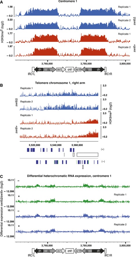
  2. H3K9me2 ChIP‐seq enrichment profiles for swi6+ and swi6∆ cells on the telomere of the right arm of chromosome 1. Two independent biological replicates for each genotype were performed (replicates 1 and 2).
  3. Differential expression of transcripts coming from centromere 1 assessed by tiling microarray gene expression analysis. Expression data for swi6+ and swi6∆ cells were taken from a previous publication (Woolcock et al, 2012).