
Figure 1. Pol II transcription is essential for nucleolar structure and function.
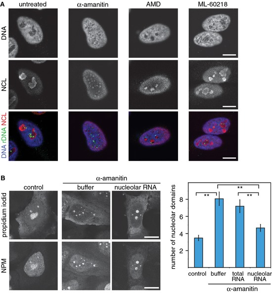
- Confocal laser scanning fluorescence microscopy (CLSM) images showing DNA staining (blue in the merged images), rDNA FISH (green) and NCL immunofluorescence (red) in untreated HeLa cells or in cells treated with α‐amanitin (50 μg/ml), AMD (50 ng/ml) or the Pol III inhibitor ML‐60218 (200 μM) for 5 h.
- CLSM images of propidium iodide‐stained RNA after microinjection of buffer or nucleolar RNA into α‐amanitin‐treated HeLa cells. Nucleoli were visualized by immunofluorescence of nucleophosmin (NPM). The graph represents the average number of nucleolar domains (± 95% CI) based on the analysis of 90, 87, 80 and 86 cells, respectively. **P‐value < 0.01, t‐test.
Data information: Scale bars, 10 μm.
See also Appendix Figs S1 and S2.