
Figure 3. aluRNA is localized to nucleoli and partially rescues α‐amanitin‐induced nucleolar dispersion.
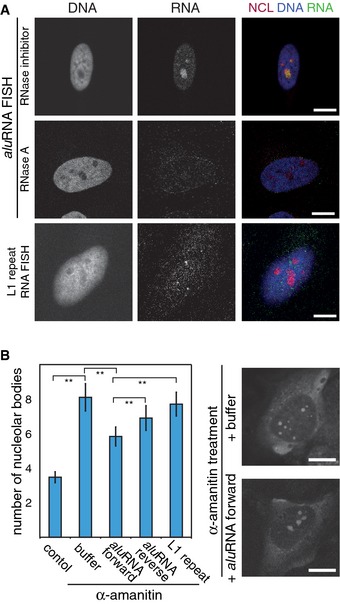
- CLSM images showing nucleolar co‐localization of NCL (immunofluorescence) with aluRNA (RNA FISH) but not with L1‐repeat RNA. Cells were pre‐treated in situ with RNase A or an RNase inhibitor. Nuclei were counterstained with DAPI. The signal intensity of nucleolar aluRNA was two‐fold higher compared to nucleoplasmic signal (n = 92, P‐value < 0.05, t‐test).
- Graphs representing the average number of nucleolar bodies after microinjection of in vitro transcribed RNA into HeLa cells that were pre‐treated with α‐amanitin (50 μg/ml) for 5 h or left untreated (control) (± 95% CI. **P‐value < 0.01, n = 90, 87, 86, 83 or 86 cells, respectively). Representative CLSM images of propidium iodide‐stained RNA are shown on the right side.
Data information: Scale bars, 10 μm.
See also Appendix Fig S5, Appendix Table S4.