Figure 2.

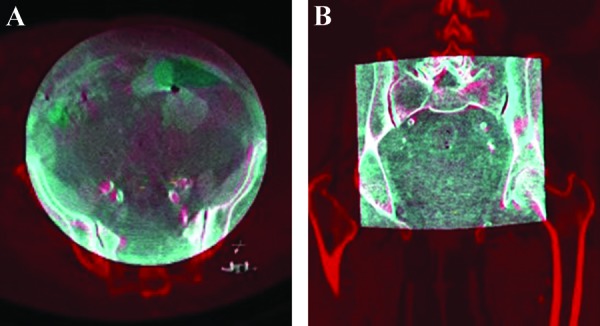
3D-3D registration in axial (A) and coronal (B) planes. The cone-beam computed tomography (CT; gray) is superimposed on the CT angiography reconstruction (red). Note the matching of bony structures and arterial calcifications.

3D-3D registration in axial (A) and coronal (B) planes. The cone-beam computed tomography (CT; gray) is superimposed on the CT angiography reconstruction (red). Note the matching of bony structures and arterial calcifications.