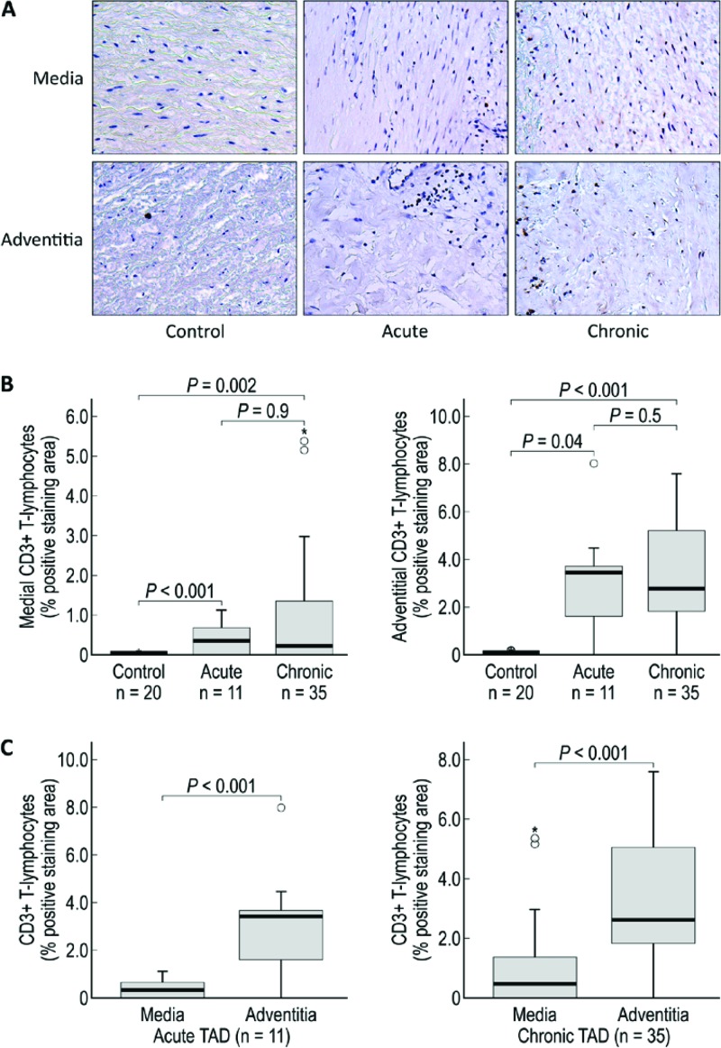
Figure 4.
CD3+ T lymphocytes are increased in the media and adventitia of acute and chronic TAD tissues. A. Immunohistochemistry staining and comparison of CD3+ T lymphocytes in the medial and adventitial layers of the aorta from donor controls and acute and chronic TAD patients. Original magnification, 200×. B. Comparison of the positive-staining areas of control, acute, and chronic dissection tissues in the media and adventitia. C. Comparison of the positive-staining areas of the media and adventitia in acute and chronic TAD. The tips of the projecting bars represent the minimum and maximum values, and the box depicts the interquartile range, with the solid middle line representing the median. Circles and asterisks represent 1.5× and 3× the interquartile range, respectively.

