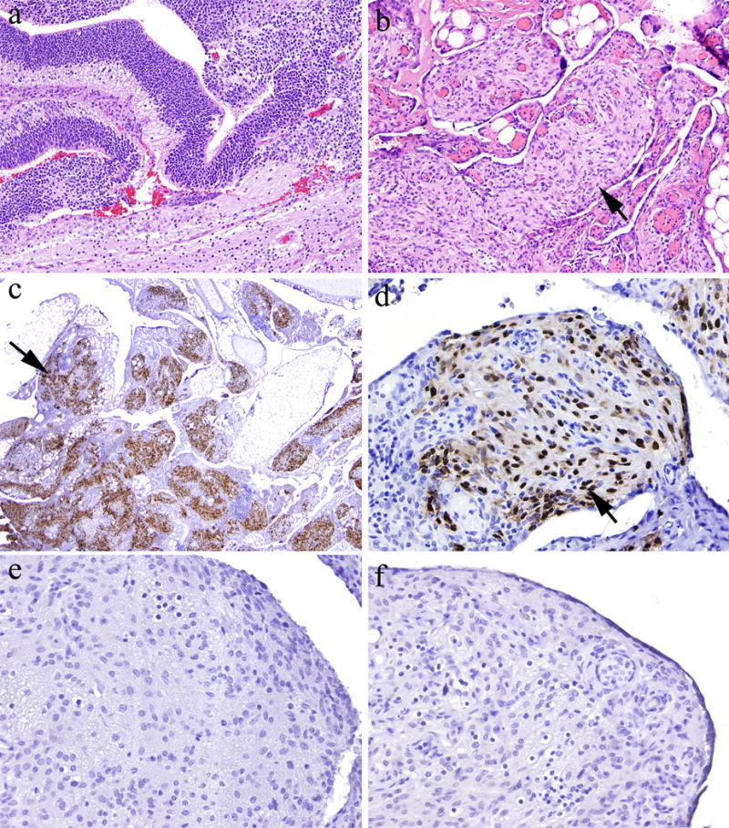
Figure 1.

Staining of gliomatosis peritonei specimens for OCT4, NANOG, and SOX2. [a] Patient’s ovarian mass was a mixed germ cell tumor, predominantly composed of immature teratoma (hematoxylin and eosin stain; original magnification, ×100). [b] Mature glial tissue in the peritoneal cavity demonstrated a micronodular growth pattern, consistent with gliomatosis peritonei (hematoxylin and eosin stain; original magnification, ×100). [c and d] Glial cells demonstrated strong, diffuse nuclear staining for SOX2 (immunohistochemical stain; original magnification, ×20 and ×200 in c and d, respectively). [e] Glial cells were negative for OCT4 (immunohistochemical stain; original magnification, ×200). [f] Glial cells were negative for NANOG (immunohistochemical stain; original magnification, ×200).
