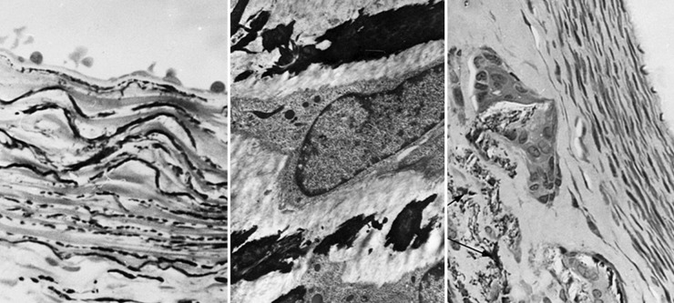
Figure 2.
Periarterial calcium chloride. Left, Light microscopic section through the rabbit right common carotid artery (RCCA) 3 days after periarterial CaCl2 showing calcium precipitation within the internal elastic lamina and elastic layers of the media (Von Kossa, 640×). Middle, Transmission electron micrograph (pyroantimonate stain) of a CCA 1 week after CaCl2 showing calcium deposits (black) within the medial elastica (4200×). Right, RCCA 6 weeks after CaCl2 application showing calcium precipitation within the media with giant cells showing central vacuolization and engulfment of fragmented calcium-elastica beneath an area of intimal proliferation (Von Kossa, 350×). Reprinted with permission from Gertz et al., J Clin Invest. 1988;81:649-656.

