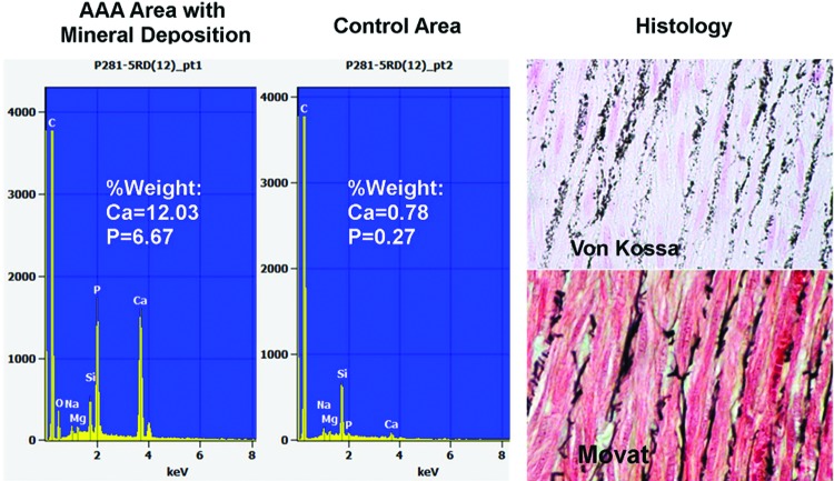
Figure 8.
Left, Elemental analysis of porcine AAA by scanning electron microscopy (SEM) with electron dispersive spectrometery (EDS) showing calcium precipitates in the elastic lamellae. Right, top = Von Kossa-stained section showing calcium precipitates within the medial elastica as additionally confirmed with Movat pentachrome (bottom) that stains elastica black.

