Figure 3.

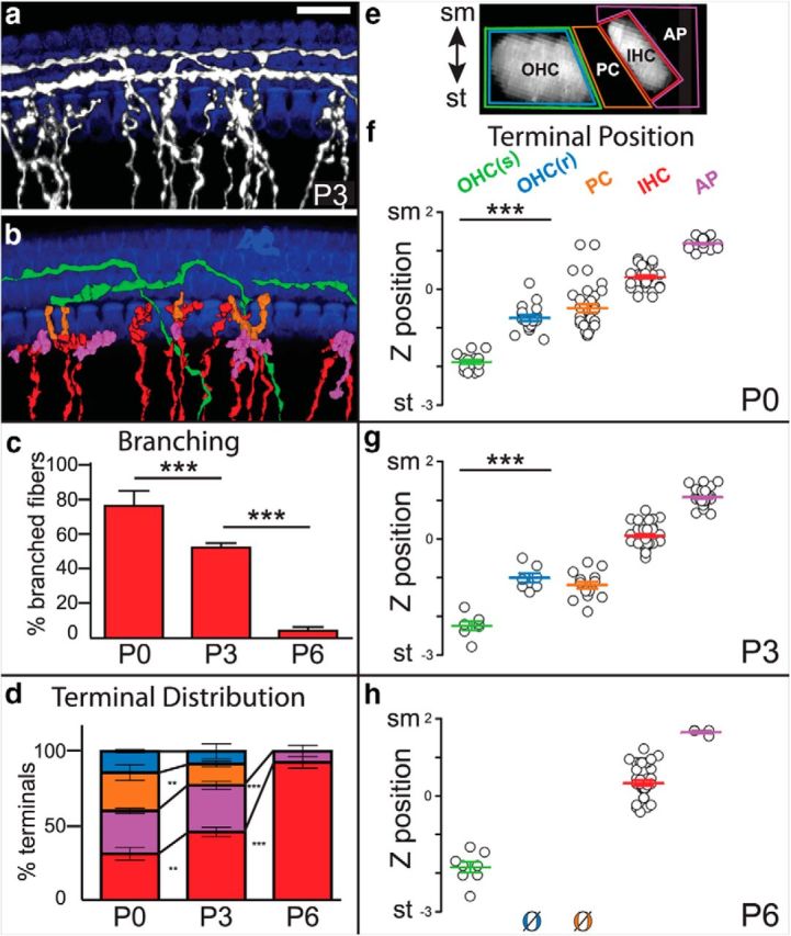
Branches from radial fibers make off-target endings that are progressively lost postnatally. a, A flat-mount view of a P3 Neurog1-CreERT2;AI14 cochlea with sparsely labeled SGNs (gray) that was immunostained for DsRed to label processes and MyoVIIa to label IHCs and OHCs (blue). b, 3D reconstructions of individual radial (red) or spiral (green) fibers were generated, and branches were color-coded according to their origin (radial or spiral) and final position, with branches associated with pillar cells (orange), IHCs (red), or instead ending just apical to the IHCs (purple). In this example, no radial fiber branches contacted an OHC. None of the spiral fibers extended branches outside of the OHC region at any age (P0: 13 spiral fibers; P3: 7 spiral fibers; P6: 8 spiral fibers). c, Quantification of branching of fibers at IHCs over time. Branching decreased significantly between P0 and P3, and between P3 and P6 (P0: 5 cochlea, 54 fibers; P3: 5 cochlea, 65 fibers; P6: 3 cochlea, 66 fibers; ***p < 0.0001, one-way ANOVA with Bonferroni correction). d, Quantification of radial terminal distribution over developmental time. At P0, a low percentage of branches contacted IHCs (red), with most branches (88/129) instead making off-target contacts apical to the hair cell (purple), with pillar cells (orange), or with OHCs in the first row (blue). The percentage of off-target endings decreased at P3 (55/104) and by P6 (4/41), all branches were confined to the IHC region, with just a handful ending in a slightly apical position. Averages of terminal distribution across cochleae are shown (P0: 4 cochleae, 129 terminals; P3: 4 cochleae, 104 terminals; P6: 3 cochleae, 41 terminals; **p < 0.005, ***p < 0.0001, one-way ANOVA with Bonferroni correction). e, A side-view of the organ of Corti showing the regions used to classify and count SGN terminals. The position of each ending was mapped and color-coded according to its proximity to an OHC (green for spiral fibers; blue for radial fibers), PC (orange), IHC (red), or apical to an IHC (purple). f–h, The z-position of each terminal was plotted along a normalized sm–st axis and grouped by type. Branches from radial fibers contacted IHCs close to sm, whereas spiral fibers contacted OHCs close to st. Off-target endings occupied intermediate positions, except for the branches that extended apically near the IHCs. Thus, the occasional branches from radial fibers that reached the first row of OHCs [OHC(r)] were significantly closer to sm than the spiral fibers. Because branches were at a similar x-y position, these differences are independent of the slight tilt of the organ of Corti. Off-target radial terminals were mostly absent by P6 (P0: 4 cochlea, 144 terminals; P3: 4 cochlea, 107 terminals; P6: 3 cochlea, 49 terminals; ***p < 0.0001, one-way ANOVA with Bonferroni correction). Scale bar: a, 20 μm. All error bars indicate SEM.
