
Figure 4.
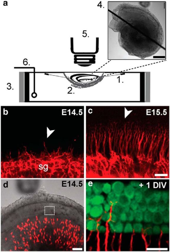
A system for imaging of intact suspended embryonic cochleae. a, A dissected cochlea is suspended with 25 μm-diameter-tungsten needles (1), cradled in a net of devascularized amniotic membrane (2), and placed in a modified dish surrounded by resistive heating elements (3). The final preparation (4) was imaged on an upright confocal microscope (5). A thin-wire thermocouple (6) monitors and maintains a physiological temperature throughout the imaging period via feedback to a temperature controller. b–e, The cochlea recapitulates normal growth and development in the chamber. In acutely dissected cochleae (b, c), SGN processes, which were labeled in bulk with tdTomato (red) by crossing Bhlhb5-cre mice to AI14 reporters, are extending from the spiral ganglion (sg) at E14.5 (b) and reach the organ of Corti target region by E15.5 (c). Arrowheads (b, c) indicate the front of process outgrowth. This pattern of outgrowth is maintained in the suspended culture preparation (d) of a Neurog1-CreERT2; AI14 cochlea, as evidenced by the presence of sparsely labeled tdTomato-positive SGN processes (e, red) among MyoVI-positive hair cells (e, green) after 1 d in vitro (+1 DIV; Movie 2). Scale bars: a (inset), 500 μm; b, c, 20 μm; d, 100 μm; e, 20 μm.