
Figure 8.
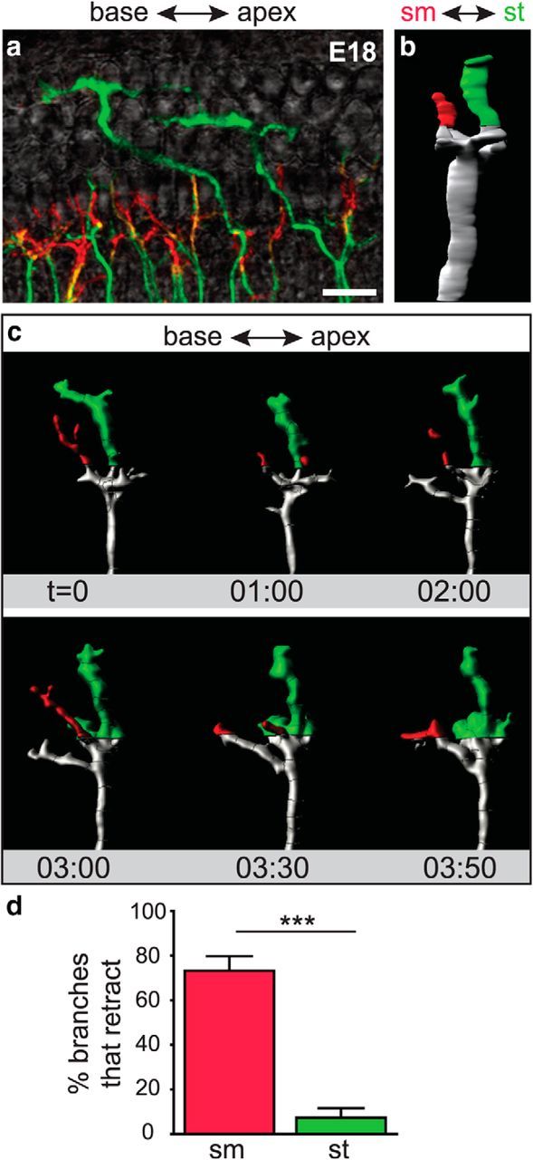
SGN peripheral processes at different depths of the organ of Corti exhibit distinct behaviors in the OHC region. a, A flat-mount view of a z-stack coded for depth illustrates the organization of SGN processes in an E18 Neurog1-CreERT2;AI14 cochlea. SGN processes in the sm half are shown in red, whereas those in the st half are green. b, A rotated view of the 3D reconstruction of an individual process, with sm to the left and st to the right. c, A montage of frames from a movie of the same reconstructed process (Movie 5), now shown in flat-mount. Branches in the OHC region were coded by depth. The branch closer to sm extended and retracted, whereas the branch positioned closer to st persisted. d, Within the OHC region, SGN processes in the sm half of the organ of Corti were significantly more likely to retract within 40 min than those in the st half. (Each region: 4 cochleae; sm region: 38 tracks; st region: 20 tracks; ***p < 0.0001, χ2 test). Scale bar, 20 μm. Error bars are SEM.