Figure 1.
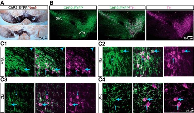
ChR2-EYFP expression in ventral midbrain dopamine neurons in DATIREScre mice after AAV-DIO-ChR2-EYFP transduction. A, Immunoperoxidase staining showing the spread of ChR2-EYFP (black) expression in the ventral midbrain with neurons visualized with NeuN (brown). With bilateral injections, ChR2-EYFP spread to both VTA and the SN in most cases (top), whereas in a minority, ChR2-EYFP spread bilaterally to the VTA but only unilaterally to the SN (bottom). B, Confocal mosaic z-projected images of the ventral midbrain showing ChR2-EYFP (green, left) and TH immunoreactivity (magenta, right). Merged image (center) shows complete overlap. C, High-power z-projected images confirming ChR2-EYFP expression in TH+ neurons in the VTA (C1), rostral linear nucleus (RLi; C2), central linear nucleus (CLi; C3), and SNc (C4). The blue arrowhead in C1 indicates the sole ChR2-EYFP+/TH− cell found in this study. Thin blue arrows indicate ChR2-EYFP−/TH+ cells and thick blue arrows indicate ChR2-EYFP+/TH+ cells.

