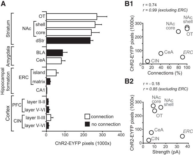
Figure 8.
Regional differences in ChR2-EYFP axon density and glutamatergic connections. A, Comparison of the density of ChR2-EYFP+ axons in the different forebrain regions expressed as the number of fluorescent pixels in the 1024 × 1024 frame (1,048,576 pixels). White bars indicate subregions where significant connections were found and black bars regions where no excitatory connections were found; n = 4 mice. B, Scatter plot showing the relationship between the incidence of connections and the density of ChR2-YFP+ axons (B1) and the strength of connections and the density of ChR2-YFP+ axons (B2). Pearson's correlation coefficients (r values) are shown for all regions with connections including and excluding the ERC.

