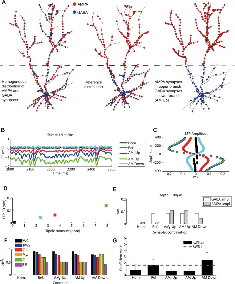
Fig 9. LFP signal and synaptic distribution.
(A) Example cases for different synaptic distributions. Left: both AMPA and GABA synapse distributed over the entire surface of the cell. Center: GABA synapses distributed only in the lower cylinder, with AMPA synapses distributed over the entire cell. Right: GABA synapses distributed only in the lower cylinder and AMPA synapses only in the upper cylinder. (B) LFP time course for different synaptic distributions: the three configurations presented in (A) correspond to black, red and green lines, respectively. Additional configurations were tested where GABA synapses were located in the lower cylinder, thalamic synapses were in both cylinders and the cortical AMPA synapses were only in the upper cylinder (blue line, AMr Up), and where all the AMPA synapses were located in the lower cylinder (cyan line, AM Down). (C) LFP amplitude as a function of depth (similar to Fig 2A) for different synaptic distributions. Blue and green markers were superimposed, illustrating that changing the position of the thalamic synapses does not alter the amplitude of the LFP (only but its mean value, cf. panel B). (D) Average LFP absolute amplitude over depths versus dipole moment (standard deviation over time) for the different synaptic distributions. (E) Contribution of AMPA and GABA currents to LFP fluctuation amplitudes for different synaptic distributions. (F) Average fraction of LFP variance explained by different proxies for different cylinder distances. Error bars are not displayed since they would not be visible in the figure. Same proxy arrangement as Fig 5C. (G) Mean and standard deviation across depths of optimal coefficients of α in the WS proxy as a function of synaptic distribution. Dashed line indicates the fixed coefficient of the RWS proxy. Note that since for homogeneous synaptic distribution the performance of the fit was low (see panel (F)), the fitted value of the relative weight of AMPA and GABA currents in contributing to the LFP signal has little significance.

